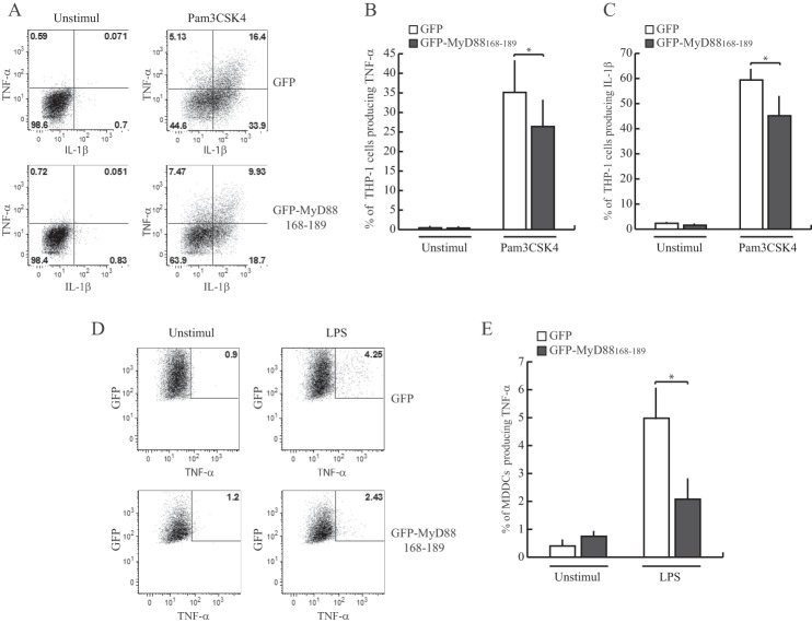
FIGURE 7.
GFP-MyD88168–189 interferes with the production of TNF-α and IL-1β induced by stimulation of TLR2 and TLR4 in human monocytoid cells. A, the pro-monocytic THP-1 cells stably expressing GFP or GFP-MyD88168–189 were differentiated into mature monocytic cells by phorbol 12-myristate 13-acetate treatment for 24 h and stimulated with Pam3CSK4 (1 μg/ml) for 6 h in presence of Golgi inhibitor brefeldin A (10 μg/ml). The percentage of TNF-α- or IL-1β-producing cells within GFP-positive cells was quantified by intracellular staining with the specific antibodies and flow cytometry analysis. B and C, bar graph represents quantitative data from three independent experiments (means ± S.E., error bars). *, p < 0.05 (paired t test). D, MDDCs transfected with constructs for transient expression of GFP or GFP-MyD88168–189 were stimulated with LPS (1 μg/ml) for 6 h in presence of Golgi inhibitor brefeldin A (10 μg/ml). At the end of incubation, the cells were harvested, and the percentage of TNF-α-producing cells within alive GFP-positive cells was quantified by flow cytometry. E, quantification of results as in D for three independent experiments was performed with cells from three individual donors. Data are represented as mean ± S.E. *, p < 0.05 (paired t test).

