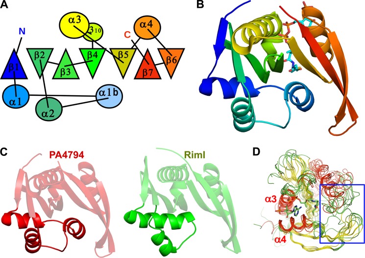
FIGURE 1.
Overall structure of PA4794 and similarity to other GNATs. A, topological diagram showing the arrangement of secondary structural elements. α-Helices are shown as circles, β-strands are shown as triangles. Orientation of the triangles shows the orientation of the β-strands, with vertex of the triangle pointing up for the β-strand pointing toward the reader, and vertex of the triangle pointing down for opposite orientation of the β-strand. B, ribbon diagram colored from N to C, Ac-CoA is shown as sticks. C, ribbon diagrams of PA4794 and RimI. Fragments showing the largest differences are highlighted. D, superposition of PA4794 and related GNATs. Conservation of the Ac-CoA/CoA binding site and the flexibility of the substrate-binding site (indicated by a blue rectangle) are visible. CoA is shown as sticks.

