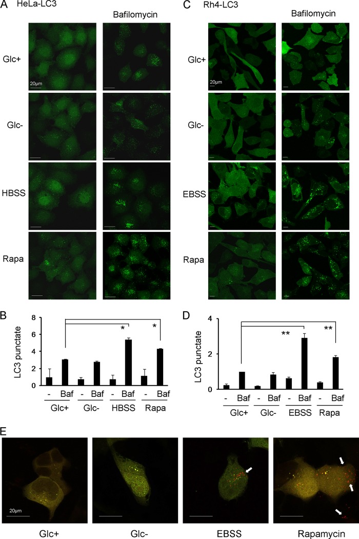
FIGURE 4.
Glucose deprivation does not induce autophagic flux. A–D, HeLa (A and B) or Rh4 (C and D) stably transfected with GFP-LC3 (HeLa-LC3, Rh4-LC3) were treated with DMEM with glucose (Glc+), glucose deprivation (Glc−), amino acid deprivation (Hank's balanced salt solution/EBSS) or rapamycin (Rapa) for 6 h with or without bafilomycin (Baf) for the last 3 h. The expression pattern of GFP-LC3 was examined under a confocal microscope. Punctate LC3 in cells was measured as described under “Experimental Procedures.” B and D show the mean + S.E. from two (HeLa, B) or four (Rh4, D) independent experiments. E, Rh4 were transfected with mRFP-GFP-LC3 plasmid and treated 24 h post-transfection for 15 h. Representative images from three independent experiments are shown. Arrows indicate red points (autophagolysosomes).

