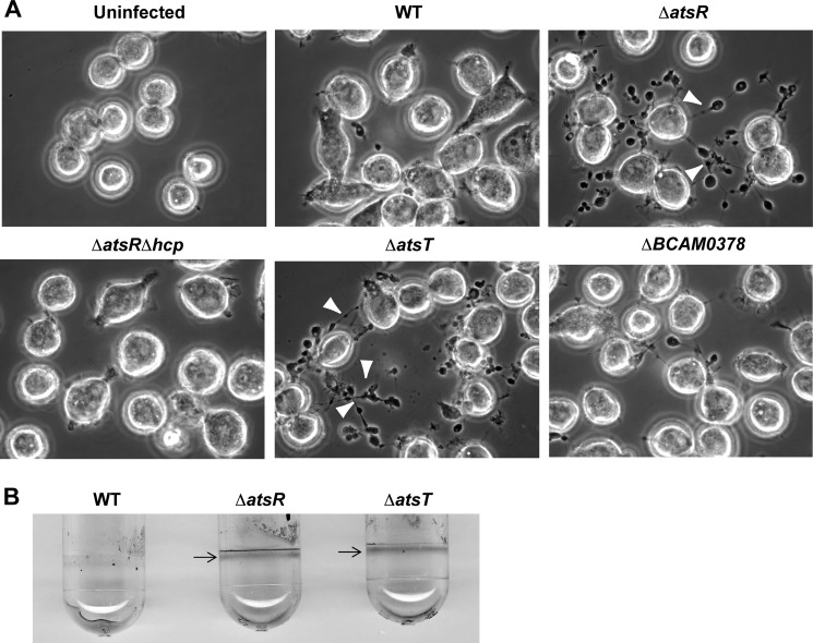
FIGURE 1.
T6SS activity and biofilm formation of B. cenocepacia K56-2 (WT) and its mutant derivatives. A, phase-contrast microscopy of infected ANA-1 murine macrophages to assess T6SS activity. Infections were performed at a multiplicity of infection of 50 for 4 h. White arrowheads indicate the presence of ectopic actin nucleation (pearls-on-a-string phenotype (15, 22)) extending from infected macrophages, which denotes T6SS activity. B. cenocepacia K56-2 ΔatsRΔhcp, a T6SS-defective mutant, was used as a negative control during the infections. Experiments consisted of three independent biological repeats where similar results were obtained. B, biofilm formation by parental strains, ΔatsR and ΔatsT mutants. B. cenocepacia K56-2 wild-type and derivative mutants were tested for biofilm formation by crystal violet staining. Arrows indicate the ring corresponding to the biofilm formation characteristic in ΔatsR (15) and ΔatsT mutants. The experiment was repeated three times in triplicate, and pictures were taken after 24 h of static incubation at 37 °C.

