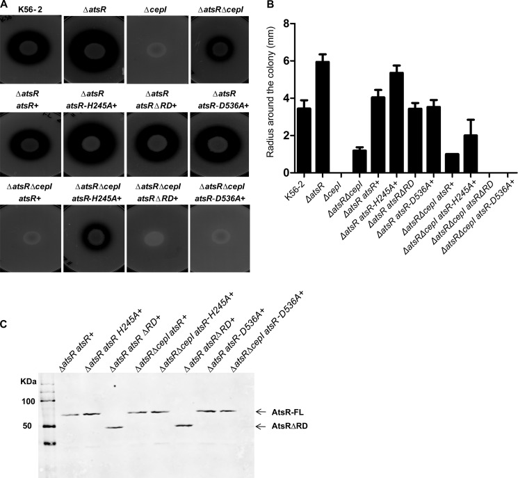
FIGURE 6.
Proteolytic activity of B. cenocepacia K56-2 wild-type, ΔatsR, ΔatsRΔcepI, and ΔcepI mutants and complemented mutants at the chromosomal level in different genetic backgrounds. A, proteolysis was tested on D-BHI milk agar plates. The plates shown are representatives of three experiments performed in triplicate. Zones of clearing around the colonies were measured at 48 h of incubation at 37 °C. B, values are the average radius ± S.D. in millimeters of three experiments performed in triplicate. C, anti-His Western blot analysis of the His-tag-purified membrane pellet of AtsR, AtsRΔRD, AtsRD536A, and AtsRH245A in B. cenocepacia. Arrows indicate the positions of full-length AtsR (AtsR, AtsRD536A, and AtsRH245A) and AtsRΔRD. AtsR-FL, full-length AtsR.

