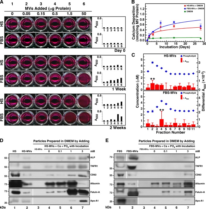
FIGURE 3.
Serum MVs induce mineral precipitation in cell culture conditions. A, MVs prepared by ultracentrifugation from HS and FBS were quantified based on total protein content prior to inoculation into DMEM at the amount indicated (Day 0, final volume of 1 ml). The solutions were incubated in cell culture conditions for the time indicated. MVs produced visible precipitation that increased in a time-dependent manner. DMEM used as a negative control produced no precipitation. B, the ability of MVs isolated by sucrose gradient centrifugation to undergo calcification. Phospholipid-rich MV fractions corresponding to 30 μg of total protein were added into DMEM and incubated in cell culture conditions. Precipitates were collected at the time indicated following centrifugation at 12,000 × g for 30 min, and the calcium content was determined using the O-cresolphthalein complexone assay. Both HS-MVs and FBS-MVs induced the formation of calcified precipitate in a time-dependent manner during incubation. C, phospholipid-rich MV fractions induce mineral precipitation in culture. 100 μl of each sucrose gradient fraction was added into DMEM in a final volume of 1 ml prior to incubation in cell culture conditions for 2 months. Mineral precipitation was assessed by A650 turbidity readings. Turbidity observed before incubation was subtracted from the turbidity reading obtained after incubation (Differential A650). The content of phospholipids was determined using a standardized biochemical assay. Phospholipid-rich fractions produced slightly higher levels of precipitation under these conditions. D and E, Western blotting analysis of co-precipitates containing MVs and mineralo-organic NPs. MVs were inoculated into DMEM, and CaCl2 and NaH2PO4 were added at the concentration indicated in a final volume of 1 ml prior to incubation in cell culture conditions for 1 day. Mineral precipitates were pelleted by centrifugation at 12,000 × g for 30 min prior to washing steps in HEPES buffer. Equal amounts (60 μg) of proteins from whole serum, MVs, or MV-NP co-precipitates were separated under denaturing and reducing conditions by SDS-PAGE and probed with the indicated antibodies. Some of the lanes shown here originated from two different gels run in parallel; these are delineated by the blank space seen in some of the blots. **, 0.1 < p < 0.5. Error bars, S.E.

