FIGURE 2.
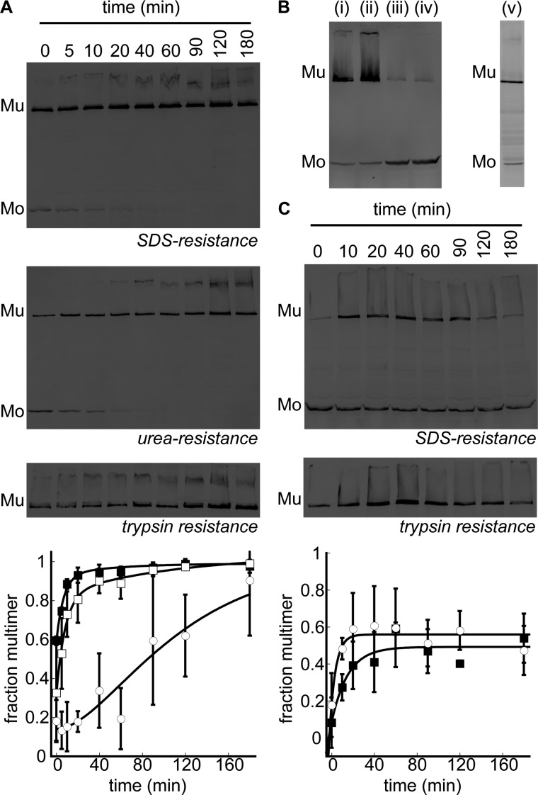
Kinetics of PulD28–42/259–660 assembly in lecithin or diC16:0PC liposomes. A, immunoblot analysis after SDS-PAGE of multimerization kinetics (■) and acquisition of urea (□) and trypsin resistance (○) of PulD28–42/259–660 in the presence of 53 mm lecithin at 30 °C. Synthesis (10 min) was performed before kinetics were monitored. Mu, multimeric PulD28–42/259–660 species; Mo, monomeric PulD28–42/259–660 species. B, PulD28–42/259–660 assembly in diCx:0PC liposomes (in i, ii, iii, and iv, x = 12, 14, 16, and 18, respectively) at 30 °C and in diC16:0PC liposomes upon warming to 42 °C (v). Synthesis was performed for 6 h, except in the latter sample, where synthesis was stopped after 10 min before increasing the temperature to 42 °C for 6 h. C, multimerization kinetics (■) and acquisition of the trypsin resistance (○) of PulD28–42/259–660 in the presence of diC16:0PC liposomes after a temperature jump from 30 to 42 °C. Synthesis was stopped after 10 min at 30 °C. Only multimers are shown for the trypsin-resistant state because monomers were digested completely. Bands were quantified by densitometry. Error bars represent S.D. over three independent measurements.

