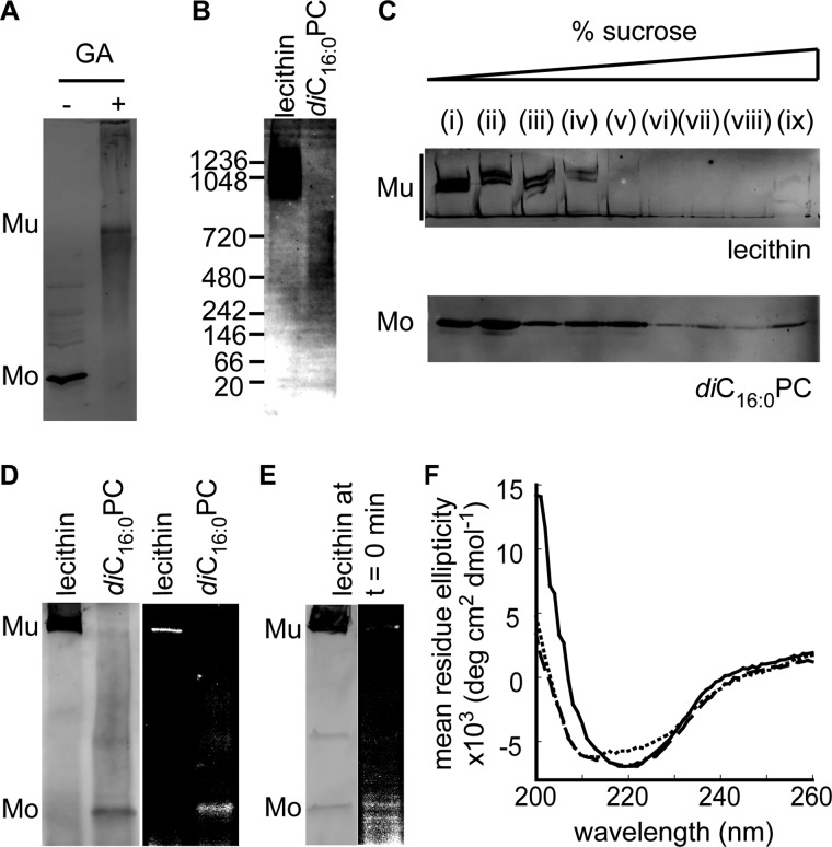
FIGURE 3.
Characterization of PulD28–42/259–66 assembled in lecithin and diC16:0PC liposomes. Shown are glutaraldehyde (GA, 100 mm) cross-linking of PulD28–42/259–660 in diC16:0PC liposomes (A) and blue native PAGE in digitonin upon membrane solubilization (B). Mu, multimeric PulD28–42/259–660 species; Mo, monomeric PulD28–42/259–660 species. C, flotation on a discontinuous sucrose gradient after assembly in liposomes. D, cross-linking of 1-azidopyrene to PulD segments inserted into the membrane. E, immunoblot analysis and 1-azidopyrene fluorescence after SDS-PAGE following a 10 min synthesis reaction in the presence of lecithin liposomes. F, circular dichroism spectra in lecithin liposomes (solid line) and in diC16:0PC liposomes before (short dashed line) and after (long dashed line) a temperature jump from 30 to 42 °C. All electrophoresed samples were analyzed by immunoblotting. All synthesis reactions were performed for 6 h at 30 °C in the presence of 4 mm liposomes, except for PulD28–42/259–660 in diC16:0PC liposomes at 42 °C, for which synthesis at 30 °C was stopped after 10 min before incubating for 6 h at 42 °C.

