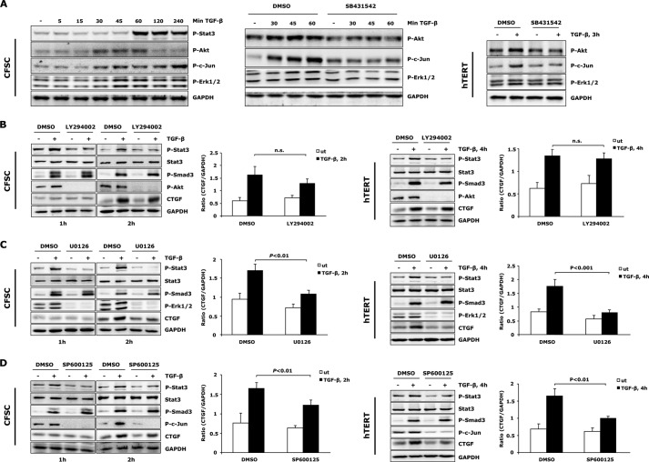
FIGURE 7.
MAPKs and PI3K are required for TGF-β-induced Stat3 activation in activated HSCs. A, Western blot revealed ALK5 (SB431542, 5 μm) mediated activation of Erk1/2, JNK, and PI3K/Akt pathways prior to Stat3 phosphorylation upon TGF-β treatment. B–D, PI3K/Akt (LY294002, 10 μm), Erk1/2 (U0126, 10 μm), and JNK (SP600125, 5 μm) inhibitors were used to block specific signaling pathways. CFSC-2G cells and hTERT HSCs were then treated with TGF-β1 (5 ng/ml) for indicated times to detect Stat3 activation and CTGF expression. All inhibitor treatments attenuated early Stat3 phosphorylation. CTGF expression was reduced by Erk1/2 and JNK inhibition but remained unchanged in the absence of PI3K. In contrast to the enhanced p-Smad3 level in LY294002 treated CFSC-2G cells (TGF-β1, 2 h), a reduced p-Smad3 level was observed in hTERT HSCs after PI3K inhibition (TGF-β1, 4 h), indicating a third signaling pathway participating in modulation of total CTGF production in this setting (see “Discussion”). CTGF and GAPDH expression was densitometrically quantified, and results are means ± S.E., p < 0.01 and p < 0.001 indicate the statistical significance. ut, untreated; DMSO, dimethyl sulfoxide.

