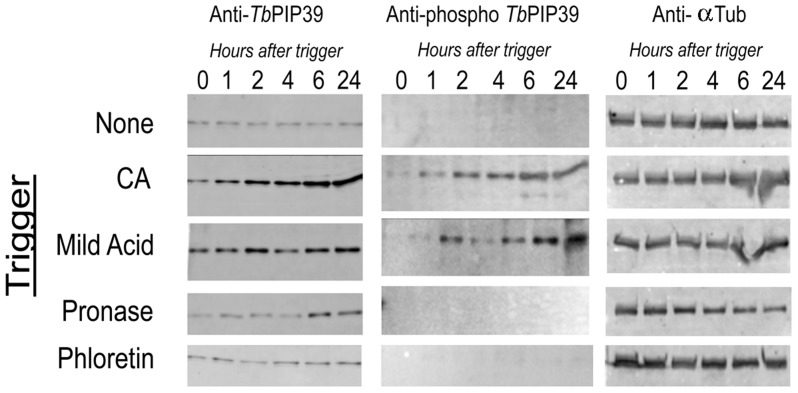
Figure 3. Differential TbPIP39 phosphorylation in response to different differentiation signals.
Stumpy cells were exposed to 6cis-aconitate (CA), mild acid (pH 5.5), pronase (4 units/ml) and phloretin (100 µM) and isolated protein samples were then reacted with a phospho-specific antibody recognising the sequence (ELDHWRTDEY*TK C) (anti-phospho TbPIP39) (middle panels). The same blot was also reacted with an anti-TbPIP39 polyclonal antibody (left hand panel) and with an antibody detecting trypanosome alpha tubulin as a loading control (right hand panels). Phosphorylated TbPIP39 was observed with CA and mild acid treatment, but not pronase.

