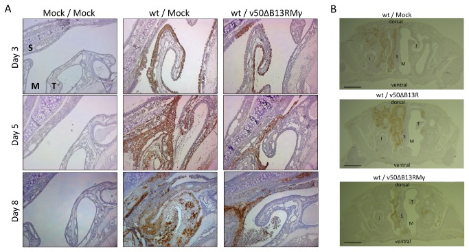
Figure 6. Histopathologic comparison and infection progression in the nasal cavity section.
Mice were infected IN with 106 pfu (~100 LD50s) of wt VACV and treated one day post-infection with 107 pfu of v50ΔB13RMγ. Mice were sacrificed at 7-8 days post-infection. All representative sections were stained with polyclonal antibodies against VACV. S=septum, T=maxilloturbinate, M=meatus (air passage), i=incisor. (A) Maxilloturbinate section, 2 mm depth, 100X magnification (B) Whole section, 5-6 mm depth. 10X magnification. Lines indicate 200 μm length.

