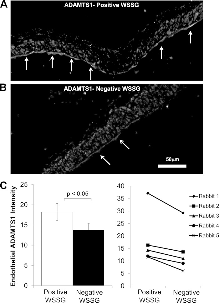
Fig. 6.
Endothelial ADAMTS1 protein is increased in the positive WSSG region compared with the negative WSS region at the basilar terminus. The basilar terminus of rabbits 5 days after carotid artery ligation was stained for ADAMTS1 by indirect immunofluorescence, and regions of positive WSSG and negative WSSG were determined by mapping computational fluid dynamics onto histology as in Fig. 7. A: under positive WSSG, the endothelial layer stained strongly for ADAMTS1 (arrows). ADAMTS1 staining was also present in many cells in the media. B: under negative WSSG, ADAMTS1 staining was weak in ECs and the other cells of the vessel. C: fluorescence intensity specifically within ECs was measured as described in the materials and methods for each of the 5 animals. Average mean EC intensity was higher in positive WSSG than in negative WSSG regions. Bars represent mean intensity ± SE from 5 animals. Intensities were significantly different between positive WSSG and negative WSSG (P < 0.05, paired t-test).

