
Fig. 5.
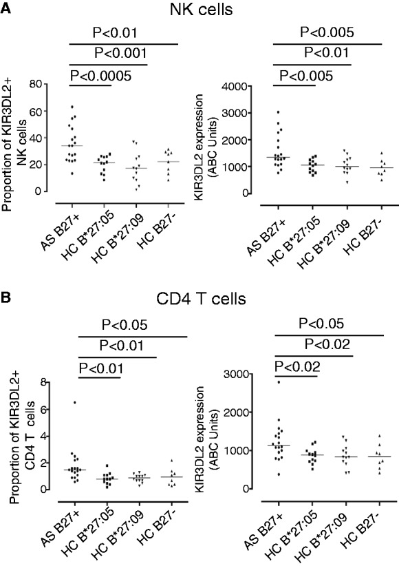
Increased expression of KIR3DL2 by leucocytes from HLA-B*27:05+ patients compared with HLA-B27− and HLA-B*27:05+ and HLA-B*27:09+ healthy controls (HCs).
(A) Left hand panel: percentage of NK cells expressing KIR3DL2 in HLA-B*27:05 + SpA patients, HLA-B*27:05+ HC, HLA-B*27:09 HC and HLA-B27 negative HC. Percentages of NK cells expressing KIR3DL2 were 35.6 ± 13.6 (mean ± 1s.d.), 20.3 ± 6.3 (P = 0.0004), 17.9 ± 11.3 (P = 0.0007) and 22.3 ± 8.5 (P = 0.007) for each of the respective groups. Right hand panel: level of expression of KIR3DL2 (ABC units) by SpA patient NK cells, HLA-B*27:05+, HLA-B*27:09+ and HLA-B27− HC. HLA-B*27:05+ SpA patients expressed 1571 ± 631.9 ABC units compared with 1038 ± 235.7 units (P = 0.003) for HLA-B*27:05 HC, 1049 ± 331.5 (P = 0.007) for HLA-B*27:09+ HC and 961 ± 318 units (P = 0.003) for HLA-B27 negative HC. (B) Left hand panel: percentages of CD4 T cells expressing KIR3DL2 in HLA-B*27:05 + SpA patients, HLA-B*27:05+, HLA-B*27:09 + and HLA-B27 negative HC. A mean of 1.8 ± 1.3 SpA CD4 T cells, 0.8 ± 0.4 HLA-B*27:05 HC (P = 0.01), 0.9 ± 0.3 B*27:09 HC (P = 0.009) and 1.0 ± 0.7 (P = 0.04) B27 negative HC expressed KIR3DL2. Right hand panel: SpA patient CD4 T cells expressed 1232 ± 513.8 ABC units of KIR3DL2, compared with 878.3 ± 199 HLA-B*27:05 HC (P = 0.011), 859.3 ± 288.7 HLA-B*27:09 HC (P = 0.016) and 847 ± 321 B27 negative HC (P = 0.030).