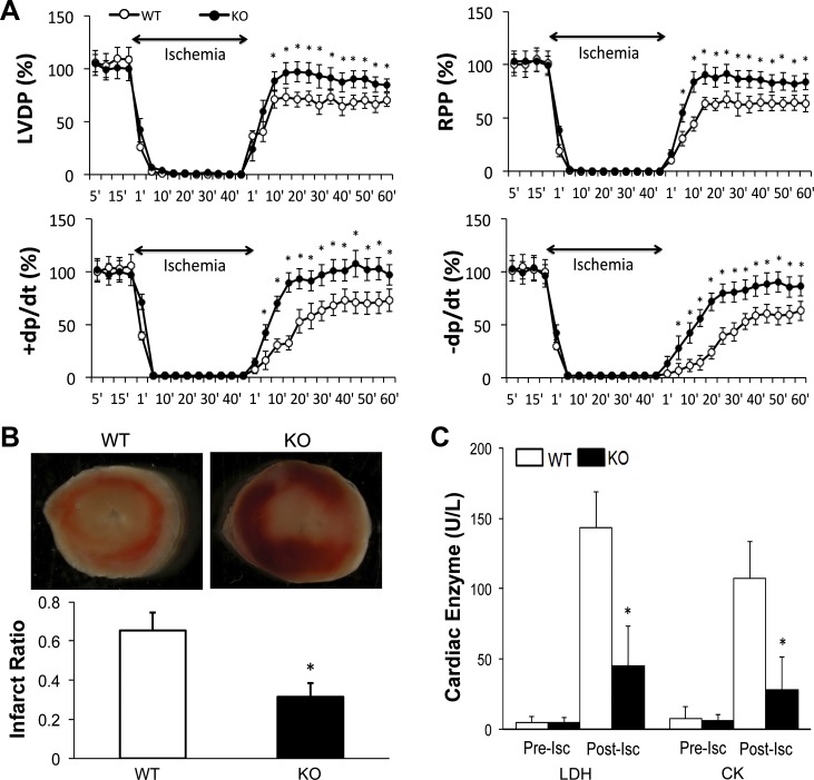
Fig. 3.
Ex vivo perfusion of WT and CMC p65-deficient mouse hearts undergoing simulated global ischemia-reperfusion. A: pressure measurements during the experimental period as a percentage of contractile function. n = 7–9/group. LVDP, left ventricular (LV) developed pressure; RPP, rate pressure product; +dP/dt, rate of rise of LV pressure per unit time; −dP/dt, rate of fall of LV pressure. B: triphenyltetrazolium chloride (TTC) staining and infarct size depicted as a ratio of infarcted area to whole heart tissue (n = 5/group). C: myocardial enzyme release for lactate dehydrogenase (LDH) and creatine phosphokinase (LDH, CK). n = 7–9. Pre-isc, preischemic; post-isc, postischemic. For each experiment, *P < 0.05, vs. WT.

