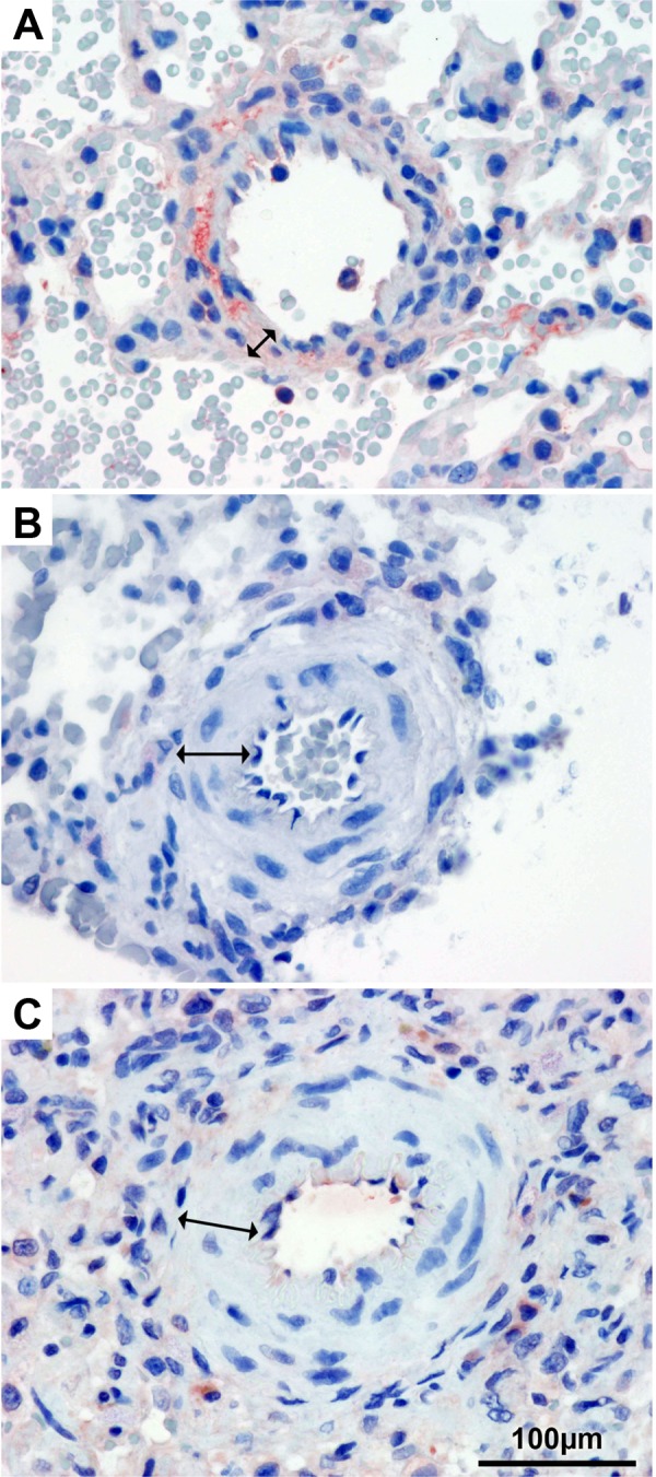
Fig. 4.

Immunohistochemistry for endothelial NO synthase. Pulmonary arteries of healthy lung tissue (A), after P/MCT (B), and after P/MCT and therapy administration (C). Arrows indicate the medial layer, which shows hypertrophy after P/MCT with and without therapy (B and C).
