
Fig. 6.
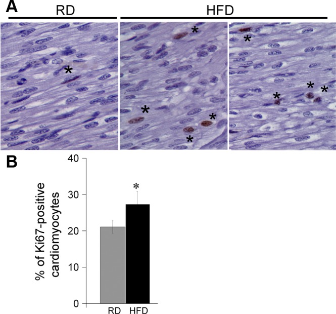
Increase in cell proliferation in HFD hearts compared with RD. Paraffin tissue sections were stained with anti-Ki-67, a marker of cell proliferation, and counterstained with DAPI. The percentage of proliferating cells was calculated by dividing the number of Ki-67-positive nuclei (*) by total nuclei and multiplying by 100. Representative images from RD and 2 HFD fetuses (A) and quantification bars (B); n = 6 for RD and 5 for HFD group. Images were analyzed using Syngene GeneTools software. Data are presented as means ± SE. *P < 0.05 (1-way ANOVA).