
Fig. 3.
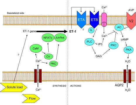
Model of ET-1 synthesis and actions in the inner medullary collecting duct. Increased tubule fluid flow increases Ca2+ entry into cells, activating calmodulin (CaM) and PKC. Ultimately, this leads to transactivation of the ET-1 promoter via NFATc and Jun/fos. Increased tubule fluid solute load also increases ET-1 synthesis, although the mechanism is unknown. ET-1 is secreted primarily basolaterally where it can activate ETA and ETB receptors in an autocrine manner. Activation of ETB leads to increased phospholipase C (PLC) activity, which increases diacylglycerol (DAG), activates PKC which inhibits vasopressin (AVP)-V2 receptor-stimulated adenylyl cyclase (AC) activity. In addition, ETB increases Ca2+ entry into cells and augments PLC production of inositol trisphosphate (IP3), leading to further increases in intracellular Ca2+ concentration; Ca2+ can inhibit AC activity as well. Inhibition of AC-dependent cAMP production leads to reduced protein kinase A (PKA)-mediated phosphorylation of aquaporin-2 (AQP2) and reduced water reabsorption. Please note that the model is drawn from studies using disparate cell types under different experimental conditions, hence this should be viewed as a conceptual overview.