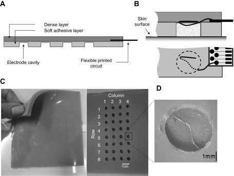
Fig. 2.
The US-EMG system of electrodes. A: schematic representation of a longitudinal section of the dense and the soft silicon layers. The layer interfacing the skin (light shaded) is soft and adhesive and houses circular cavities where wire electrodes are exposed (detailed in B and D). The external layer (dark shaded) is dense and provides sufficiently rigid support for the printed circuits connecting the electrodes to the amplifier. Pictures of the dense layer, of the adhesive layer housing electrodes, and of a single cavity containing an exposed wire electrode are shown in C and D.

