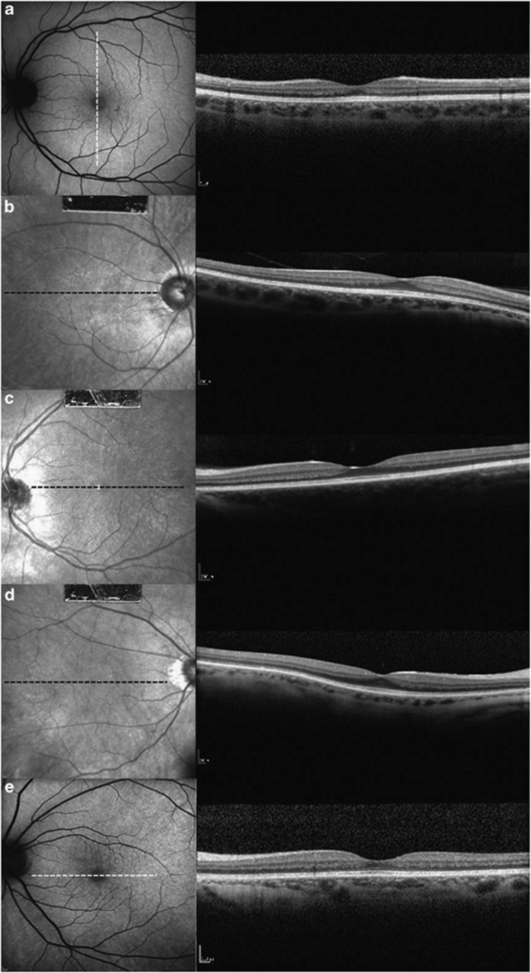
Figure 2.
High-resolution SD-OCT scans through the fovea of the five XLRP carriers. SD-OCT images of (a) subject 1, (b) subject 2, (c) subject 3, (d) subject 4 and (e) subject 5. All carriers with TLR have normal retinal microstructure and thickness on SD-OCT images. These SD-OCT images also showed an intact inner ellipsoid band, indicating the presence of photoreceptor IS/OS junctions. Photoreceptor OSs, RPE and Bruch's membrane were also intact in all carriers. The position of the SD-OCT line scan is indicated by the white or black dashed lines in AF or IR images. AF, autofluorescence; RPE, retinal pigment epithelium; SD-OCT, spectral domain optical coherence tomography.

