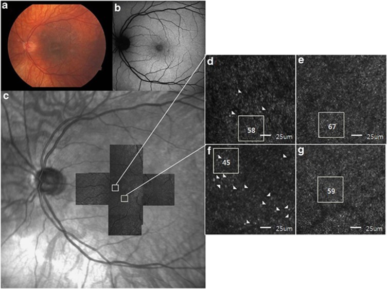
Figure 3.
Retinal images of the left eye of subject 1. (a) Fundus photograph shows fundus TLR within the posterior pole, which is more prominent temporal to the macula, and myopic changes with prominence of the choroidal vascular pattern. (b) No abnormal AF is detected on FAF imaging. (c) A montage of AO-SLO images from subject 1 matched with the IR images. (d) A magnified AO-SLO image of the area (nasal 0.5 mm) indicated by the white box in the image of c, showing a relatively regular cone cell appearance. Some morphologically large and/or dysmorphic cone cells are indicated by arrowheads, but it is not that much compared with another area (inferior 0.5 mm) in the same patient. The number of cone cells within the area indicated by the white box was estimated to be 58. (e) A magnified AO-SLO image from an unaffected control at the same location. (f) A magnified AO-SLO image of the area (inferior 0.5 mm) indicated by the white box in the image of c, shows less compact distributions than those with normal controls. Some cone cells show irregular appearance and are of variable asymmetrical sizes and shapes, which are indicated by arrowheads and the cone count is 45 in the white box area. (g) A magnified AO-SLO image from an unaffected control at the same location.

