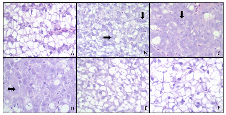
Figure 1.
Liver histopathology in AFB1-exposed red drum. Liver sections were stained with hematoxylin and eosin. Treatments were as follows: (A) 0 ppm AFB1 (B) 1 ppm AFB1 (C) 3 ppm (D) 5 ppm AFB1 (E) AFB1 + 1% NS and (F) 5 ppm AFB1 + 2% NS. Marked pleomorphism, megalokaryosis with prominent nucleoli (arrows) and loss of hepatocellular cytoplasmic macrovacuolation was observed in the treatment groups that received large amounts of aflatoxin (B,C,D). Although not significant, inclusion of NS resulted in decreased histopathological scores attributable to increased cytoplasmic vacuolation and reduced cellular pleomorphism.

