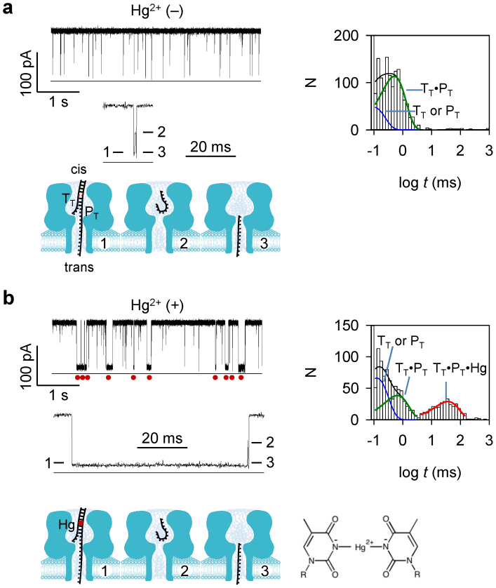
Figure 1. Detection of a single T-Hg-T MercuLock in the nanopore.
The mixture of target TT and probe PT were presented in cis solution. (a) and (b). Representative current traces, multi-level signature blocks, block duration histograms and corresponding diagram of molecular configurations, in the absence of Hg2+ (a) and in the presence of Hg2+ (b). The sequences of TT and PT are shown in Table S1. Traces were recorded at +130 mV (cis grounded) in 1 M KCl buffered with 10 mM Tris (pH 7.4). cis solution contained 1 μM TT and 1 μM PT. In b, 10 μM HgCl2 was presented in cis solution. Red dots under the trace in panel b mark the long block signatures for the TT·PT hybrid bound with a Hg2+ ion to the T-T mismatch. Values of block duration were given in Table S2. Red dot in the model in panel b represents the MercuLock formed in the DNA duplex.

