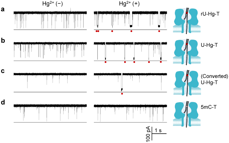
Figure 2. Discrimination of uracil and unmethylated cytosine with MercuLock.
(a) through (d) crrent trace showing signature blocks produced by various targe⃛probe hybrids TrU·PT (a), TU·PT (b), TC→U·PT (c) and TmC·PT (d) in the absence (left panel) and in the presence of Hg2+ (right panel). These hybrids contained a mismatch of uracil (uridine)-thymine (rU-T), uracil (deoxyuridine)-thymine mismatch (U-T), converted uracil-thymine (U-T), and 5-methyl cytosine-thymine (mC-T), respectively. TC→U was converted from target TC by bisulfite. Red dots under the traces marked the signature blocks for Hg2+ binding to the corresponding mismatches. Red dots in models represented the MercuLock formed in the DNA duplex. The sequences of targets TrU, TU, TC, TmC and probe PT were shown in Table S1. Traces were recorded at +130 mV in 1 M KCl solution buffered with 10 mM Tris (pH 7.4). cis solution contained 1 μM target DNAs and 1 μM PT, and 10 μM HgCl2 (right traces). The traces for TC·PT with and without Hg2+ were shown in Fig. S1. Values of block duration were given in Table S2.

