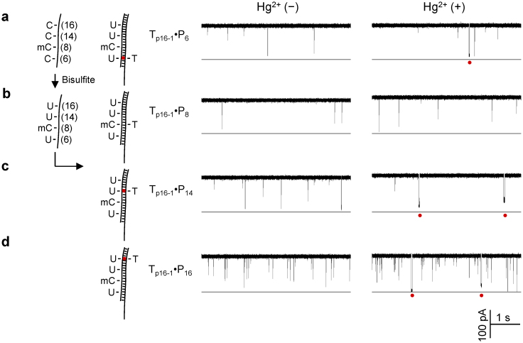
Figure 3. Site-specific detection of DNA methylation with a MercuLock.
(a) through (d) were current traces for the bisufite-converted Tp16-1 hybridized with probes PC6 (a), PC8 (b), PC14 (c) and PC16 (d) in the absence of Hg2+ (left panel) and in the presence of Hg2+ (right panel ). The four probes were designed for detecting CpG cytosines at the positions C6, C8, C14 and C16. C8 was 5-methyl cytosine (mC) and remained unchanged after bisulfite treatment. The other three positions were unmethylated cytosine (C), and thus converted to uracil (U) by bisulfite treatment. Red dots under the traces mark the signature long blocks for Hg2+ ion binding to the U-T mismatches. Red dots in the models (left) marked the MercuLock in the DNA duplex.

