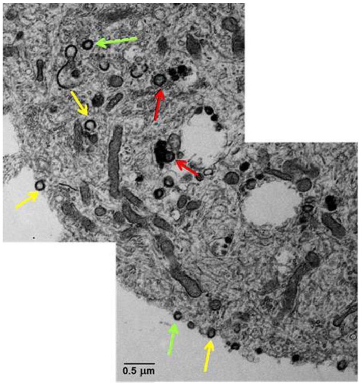
Figure 2.
Transmission electron microscopy of VZV-infected human neurons derived from induced pluripotent stem cells. A montage of the cytoplasm and cell surface of a VZV-infected neuron showed viral particles without capsids and viral DNA (yellow arrows), viral particles with capsids but not viral DNA (green arrows), and complete viral particles with capsid and DNA (red arrows). Copied and modified with permission from J. Virol [34].

