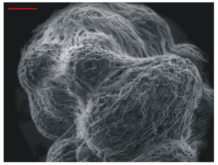
Figure 3.
Scanning electron microscopy of 3-dimensional tissue-like assemblies of normal human neuronal progenitor cells maintained for 6 months in suspension. Note the indistinguishable nature of individual cells. Cells assemble around the spherical support matrix, and multiple cell-coated matrixes fuse to form tissue-like assemblies. Bar = 100 mm. Image copied with permission from PLoS Pathogens [35].

