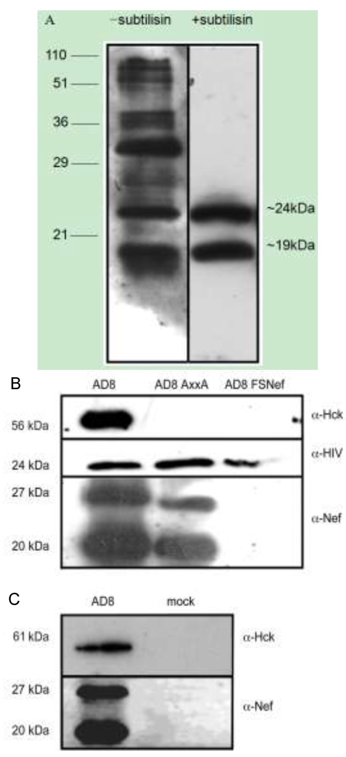
Figure 3.
Nef and Hck are present in HIV-1 virions derived from 293T cell lines and primary monocyte-derived macrophages. (A) Immunoblots of lysates of pelleted viral material generated by transfection of 293T cells with pAD8-1, with and without subtilisin digestion, probed with HIV antisera. The molecular weights of standard markers are indicated in kDa on the left-hand side of the figure. The molecular weights on the right hand side of the figure represent the approximate size of two major reactive products which likely correspond to viral capsid and nucleocapsid proteins; (B) HIV-1 viral particles purified by subtilisin treatment of pelleted virus isolated from the supernatants of 293Ts co-transfected with pAD8-1, pAD8AxxA or pAD8FSNef and pHck; or (C) of monocyte-derived macrophages infected with AD8 or mock-infected; were subjected to SDS-PAGE and probed with anti-Hck and EH1 anti-Nef mouse monoclonal sera. Data are representative from 2 independent experiments for each cell type.

