
Fig. 1.
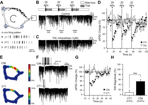
In vivo firing patterns induce depolarization-induced suppression of inhibition (DSI) in vitro. A, top: scheme representing the position at which action potentials (APs) were emitted by a CA1 place cell during 3 consecutive trials when evaluating spatial memory on a cheeseboard maze (Dupret et al. 2010). During learning, the animal is given consecutive trials to locate 3 hidden food rewards (blue circles). The start box is opened for each trial, and the animal harvests the 3 rewards before returning to the start box and collects a 4th pellet. Each dot corresponds to the location at which place cell emitted an individual spike and is superimposed on the animal's path. A, bottom: the bars represent the time at which APs were emitted by a CA1 place cell during the 1st, 2nd, and 3rd trial. B: voltage-clamp recordings [holding membrane potential (Vhold) = −80 mV] of spontaneous GABAergic activity interrupted by transient current-clamp switches during which brief direct-current (DC) injections were performed to replay the whole sequence of APs that were recorded in vivo (including the time spent in the start box). Above and with a similar time scale are depicted the time spent in the start box and the cheeseboard maze during the 3 consecutive trials together with the numbers of APs emitted during the crossing of the place field at a time depicted by an arrowhead. C: same experiment as in B except that the cell was dialyzed with tetrahydrolipstatin (THL; 1 μM). D: time course of spontaneous GABAergic charge during the replay of the whole AP sequence depicted in B and C in control (CTL; black, n = 3) and THL (1 μM, white, n = 4). The time spent in the place field is denoted in gray. sIPSC, spontaneous inhibitory postsynaptic current. E: color-coded rate map of simultaneously recorded place cells for the last 20 trials starting from those depicted in B for clusters C56 (top) and C71 (bottom). C56 is the same place cell as the 1 depicted in A. F: voltage-clamp recordings (Vhold = −80 mV) of spontaneous GABAergic activity interrupted by transient current-clamp switches during which brief DC injections were performed to replay a sequence of APs that were recorded in vivo during a single trial in 2 different place cells (C56 and C71). The insets represent the sequence of APs that was evoked in the in vitro recorded cells. G: mean time course of spontaneous GABAergic charge before and after the replay of the different AP sequences depicted in F (C56, n = 6; C71, n = 4). H: DSI magnitude measured in the next 4 s following AP discharge for the cells depicted in G. ***P < 0.001.