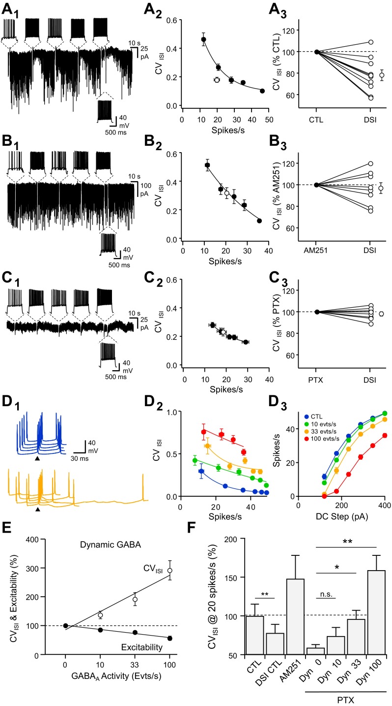
Fig. 3.
Endocannabinoid-mediated self-tuning of spike timing in pyramidal neurons. A1: voltage-clamp recordings (Vhold = −80 mV) of spontaneous GABAergic activity interrupted by episodic current-clamp switches, during which 1-s duration DC injections were performed to evoke a series of APs at various rates (top insets). The last series of APs was performed 4 s after the previous 1 to evaluate spike timing during DSI. A2: CVISI vs. spike rate for the cell depicted in A1 in control (black) and during DSI (white). A3: summary of changes in CVISI for the discharge rate during DSI vs. an equivalent rate of discharge during the control period (n = 10). B: same experiments as in A but in the presence of AM251 (5 μM, n = 8). C: same experiments as in A but in the presence of picrotoxin (PTX; 100 μM, n = 8). D1: waterfall view of membrane-potential (Vm) fluctuations from the 4th to the 5th spike of the discharge in response to a DC step (1 s, 150 pA) in the absence (blue) and when spontaneous inhibitory postsynaptic conductances (SP-IPSGs) were dynamically injected at a rate of 33 events per second (orange). The 5th spike of each discharge was set as the time reference (arrowheads). D: CVISI vs. spike firing rate (D2) and firing rate vs. DC step (D3) in control (blue) and in the presence of SP-IPSGs randomly injected at a rate of 10 (green), 33 (orange), or 100 (red) events per second (evts/s). E: changes in excitability (black) and CVISI (white) when neurons received different rates of SP-IPSGs. F: summary of changes in CVISI for a discharge rate of 20 spikes per second in control conditions (n = 10), after DSI, in the presence of AM251 (5 μM, n = 8) and in the presence of PTX and SP-IPSGs injected dynamically (Dyn) at a rate of 0 (n = 8), 10, 33, and 100 events per second (n = 5). *P < 0.05, **P < 0.01.

