Figure 2.
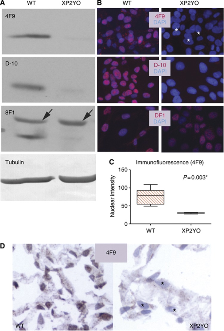
The 4F9 antibody is specific for ERCC1. Specificity of 4F9 is assessed in human skin fibroblasts isolated from either a normal individual (WT) or an individual with a mutation in XPF causing near-undetectable ERCC1 (XP2YO). (A) 4F9 is specific by western blot. Only a trace amount of ERCC1 is detected in XP2YO cells with either 4F9 or the specific anti-ERCC1 antibody D-10. In contrast, the non-specific 8F1 antibody recognises an additional band migrating slightly slower than ERCC1, present both in WT and ERCC1 deficient cells (arrows). Tubulin (loading control). (B) 4F9 is specific by immunofluorescence. Only background nuclear signal is observed in ERCC1-deficient cells either with 4F9 (white asterisks) or with the antibody D-10 while nuclear staining is readily observable in WT cells. In contrast, the nuclear signal persists in ERCC1 deficient cells when 8F1 is used, confirming the lack of specificity of this antibody. ERCC1 antibodies (red); DNA stain DAPI (blue). (C) Quantitation of average nuclear fluorescence intensity represented by boxplot; p (paired t-test); * indicates statistical significance. (D) 4F9 is specific by immunohistochemistry performed on formalin-fixed paraffin-embedded cells. Only background staining is observed in ERCC1-deficient cells (black asterisks). 4F9 (brown); haematoxylin counterstain (blue).

