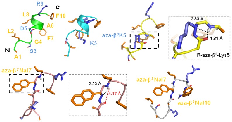
Figure 5.

The best structures (with the lowest energies) of AD (green), AK (cyan), Aβ3K (yellow), K-2Nal7 (pink) and K-2Nal (grey). The side chains of the polar and hydrophobic residues are respectively in blue and orange. Aza-β3 residues snapshots are available for the Aβ3K and K-2Nal7 peptides (see also Table S2).
