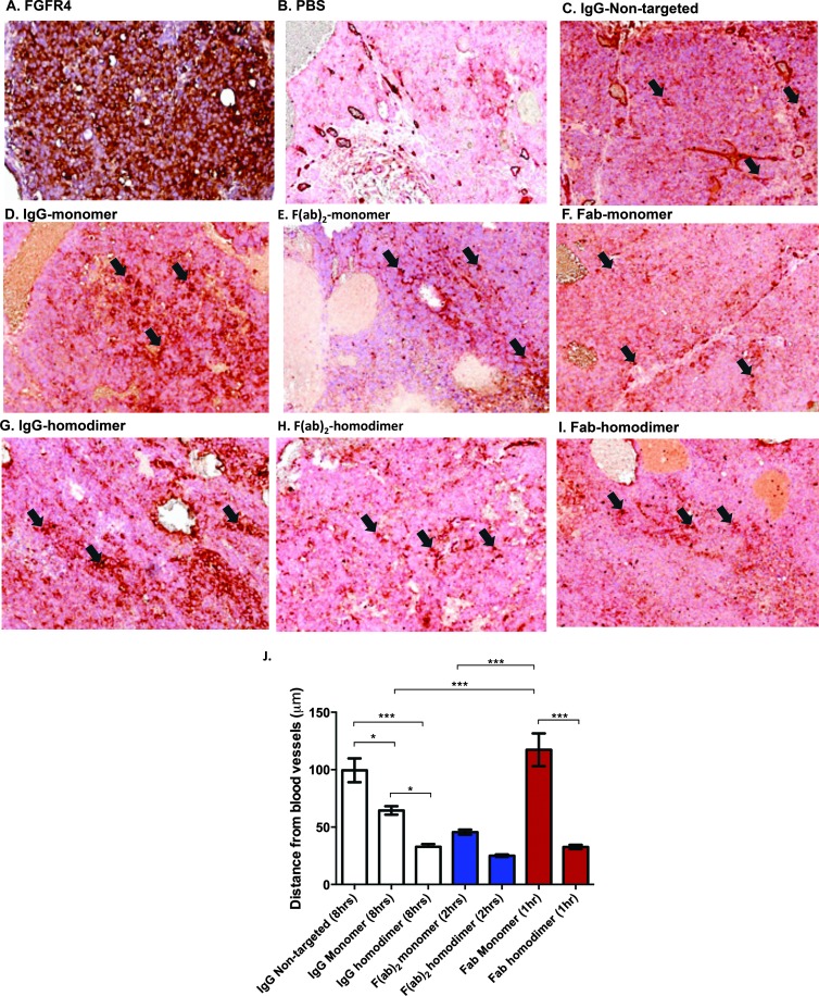
Figure 4.
Increased avidity decreases penetration of scaffolds into the tumor. (A) FGFR4 staining in an adjacent section is shown. Dual staining for blood vessels (brick red) and human IgG (brown) is shown in (B) PBS-dosed animals, (C) nontargeted IgG (8 hours), monomer peptide-conjugated (D) IgG (8 hours), (E) F(ab)2 (2 hours), (F) Fab (1 hour) and homodimer peptide-conjugated (G) IgG (8 hours), (H) F(ab)2 (2 hours) and (I) Fab (1 hour). Arrows indicate blood vessels (red). Perivascular or diffuse construct staining from those points can be seen. (J) Plot of average distance from randomly selected blood vessels (mean ± SEM; *P < .05, ***P < .001 by one-way ANOVA with Bonferroni post-test).

