
Figure 6.
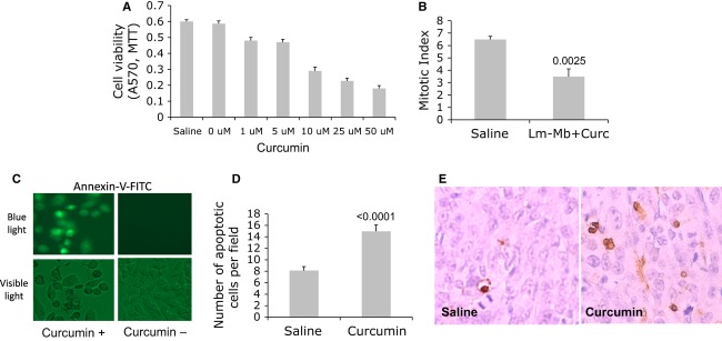
Curcumin inhibited proliferation and killed 4T1 tumor cells through apoptosis. 4T1 tumor cells were cultured with different doses of curcumin for 72 h, and cell viability was analyzed by MTT (A). We also analyzed the Mitotic Index in tumors of mice that received curcumin or saline (B). 4T1 tumor cells were cultured with 100 μmol/L of curcumin in vitro for 24 h, and subsequently incubated with anti-Annexin-V antibodies for the detection of early apoptosis (C). Primary tumors of mice that received curcumin or saline (according Immunization protocol B) were analyzed for the detection of late apoptosis in vivo by the TUNEL assay (D). Apoptotic cells in the primary tumor by the TUNEL assay and light microscopy are shown in (E). Representative of two experiments in A, C, D. Average of two experiments in B and D. n = 5 mice per group. Unpaired t test P < 0.05 is significant. Magnification light microscopy in C and E is 400×. In A, curcumin was dissolved in DMSO and then diluted to the final concentrations of 1–50 μmol/L. The 0 μmol/L represents DMSO without curcumin.