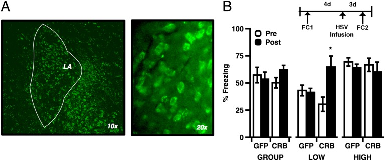
Fig. 6.
(A) GFP fluorescence from HSV-infected neurons. Cell counts estimated from samples of HSV-infected tissue indicate that 11.7% of basal/lateral amygdala cells were GFP(+) relative to DAPI. (B) Viral expression of CREBY134F selectively enhanced L-LTM memory (ANOVA, phenotype: F(2,19) = 15.604, P < 0.001; interaction: (session) F(1,20) = 32.556, P < 0.001; (construct) F(1,20) = 4.599, P = 0.044; post hoc, Low vs. Middle/High, Pre- vs. Postinfusion: t (9) = −3.788, P = 0.004). L-LTM behavior was not affected by HSV-GFP infusion (post hoc, Low vs. Middle/High, Pre- vs. Postinfusion: t (11) = −4.326, P = 0.001). Error bars indicate ± SEM; *P < 0.05.

