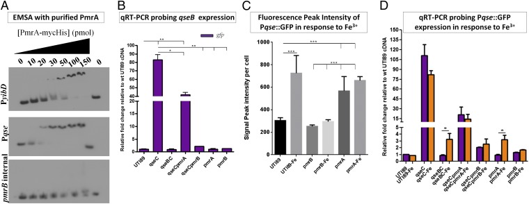
Fig. 3.
QseBC and PmrAB are physiologically interconnected. (A) Electromobility shift assays (EMSA) with purified PmrA and the qseBC promoter. The yibD promoter is a known PmrA direct target and was used as a positive control (17); an internal region of the pmrB gene was amplified and used as a negative control for binding. (B) Relative-fold change of gfp driven by the qseBC promoter in WT UTI89, UTI89ΔqseC, UTI89ΔqseBC, UTI89ΔqseCΔpmrA, UTI89ΔqseCΔpmrB, UTI89ΔpmrA, and UTI89ΔpmrB measured by qRT-PCR. (C) Graph depicting GFP intensity in bacterial cells harboring PqseBC::GFP in the presence of ferric iron, the PmrAB activating signal. Peak intensity measurements were taken for 50 bacterial cells per strain from three independent microscopy experiments. Error graphs in both graphs indicate the geometric mean with 95% confidence interval. Statistical analysis was performed using two-tailed unpaired t test, where ***P < 0.0001. (D) Relative-fold change of gfp driven by the qseBC promoter in response to Fe3+ measured by qPCR.

