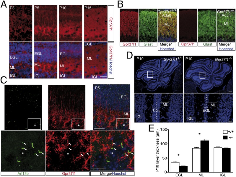
Fig. 1.
Gpr37l1 expression and cerebellar morphology of adult and developing Gpr37l1+/+ and Gpr37l1−/− male mice. (A) Cerebellar immunofluorescence labeling of Gpr37l1 and nuclear Hoechst staining in P3, P5, P10, and P15 pups and (B) immunofluorescence labeling of Gpr37l1 (red) and Glast (green) in Gpr37l1+/+ and Gpr37l1−/− adult mice. (C) Double immunofluorescence staining of Gpr37l1 and Arl13b in sagittal cerebellar sections of P5 wild-type pups. (D) Nuclear Hoechst staining in P10 cerebella of Gpr37l1+/+ and Gpr37l1−/− pups. (E) Quantification of the average thickness of the EGL, ML, and IGL in the two genotype groups at P10 (mean ± SEM, n = 3 per group). *P < 0.05 +/+ vs. −/−, unpaired t test. (Scale bars in A–C, Upper, and D, Lower, 25 μm and in C, Lower, 10 μm and D, Upper, 250 μm.) EGL, external granular layer; IGL, internal granular layer; ML, molecular layer. Asterisks indicate Purkinje neuron somata; arrows point to Gpr37l1 and Arl13b doubly positive primary cilium structures.

