Fig. 2.
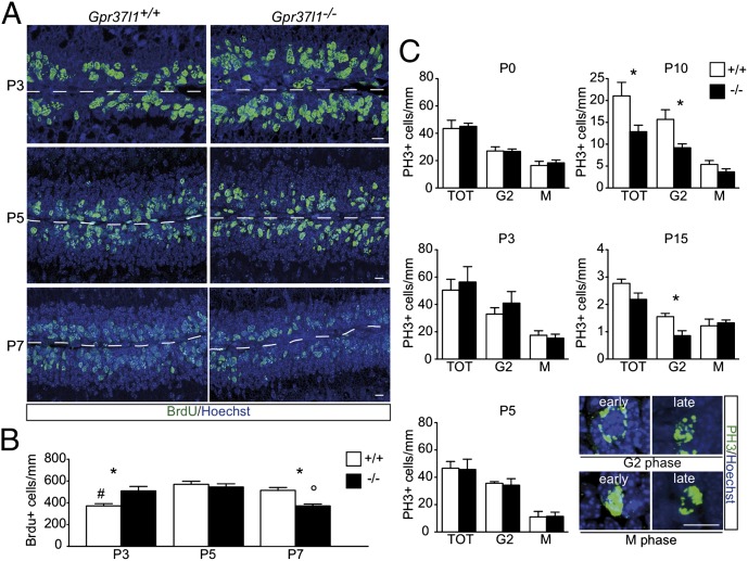
Postnatal proliferation of cerebellar granule neuron precursors in Gpr37l1+/+ and Gpr37l1−/− male mice. (A) Immunofluorescence labeling of BrdU and Hoechst staining of EGL cells at P3, P5, and P7 in Gpr37l1+/+ and Gpr37l1−/− littermates. (B) Quantification of the average number of BrdU-positive cells normalized by the EGL length (mean ± SEM), *P < 0.05 +/+ vs. −/−; #P < 0.05 P3 vs. P5, P7 in Gpr37l1+/+ mice; °P < 0.05 P7 vs. P3, P5 in Gpr37l1−/− mice, unpaired t test. (C) Quantification of the average number of phosphohistone H3 (PH3)-positive and G2- or M-phase cells at P0, P3, P5, P10, and P15 in the EGL of Gpr37l1+/+ and Gpr37l1−/− littermates (mean ± SEM n = 3 per group), *P ≤ 0.05 +/+ vs. −/−, unpaired t test. (Lower) Representative images of anti-PH3 immunofluorescence labeling and nuclear Hoechst counterstaining of EGL cells in P10 Gpr37l1+/+ pups, showing distinct PH3-positive cells in either early or late G2 phase and either early or late (mitotic division) M phase. (Scale bars in A and C, 10 μm.)

