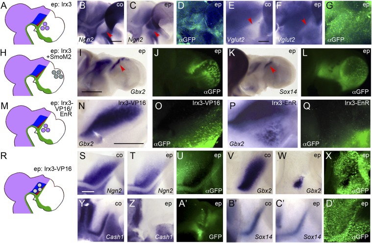
Fig. 3.
Irx3 endows cells with thalamic competence in response to Shh signaling. (A, H, M, and R) Schematic representations of electroporation experiments. Lateral views of hemisected (B–G, N–Q, and S–D’) or whole mount (I–L) chick brains electroporated (ep) between E2.5 and E3 with Irx3 into the prethalamus (B–G), with Irx3+SmoM2 into the telencephalon (I–L), with Irx3-VP16 into the prethalamus (N and O), with Irx3-EnR into the prethalamus (P and Q), or with Irx3-VP16 into the thalamus (S–D’) and fixed at 2 dpe (B–D, S–U, and Y–A’) or 3 dpe (E–G, I–L, N–Q, V–X, and B’–D’). Anterior points to the right. B, E, S, V, Y, and B’ are unelectroporated control halves (co). ISH for Ngn2 (B, C, S, and T), Vglut2 (E and F), Gbx2 (I, N, P, V, and W), Sox14 (K, B’, and C’), and Cash1 (Y and Z). D, G, J , L , O , Q , U , X , and D’ show anti-GFP immunofluorescence (αGFP); A’ shows pre-in situ GFP fluorescence. [Scale bars in B (for B–D), E (for E–G), and S (for S–D’), 0.25 mm; scale bar in I (for I–L), 1 mm; scale bar in N (for N–Q), 0.5 mm. Red arrowheads point at prethalamus in B, C, E, and F and at domains of ectopic gene expression in I and K.

