Fig. 5.
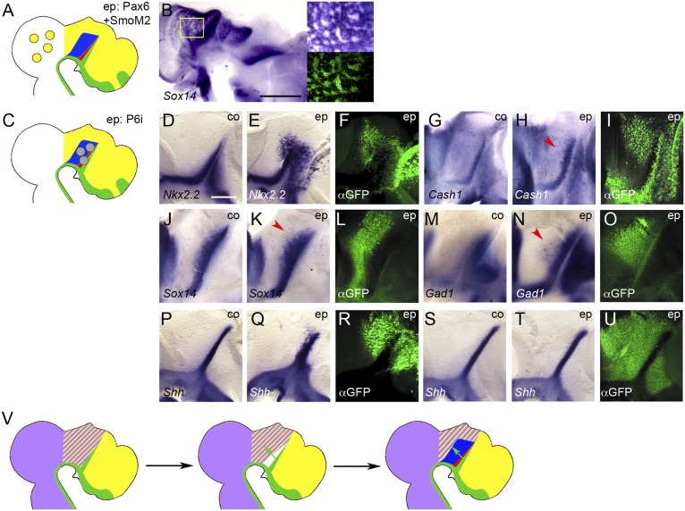
Pax6 antagonizes rTh formation. (A and C) Schematic representations of electroporation experiments. (B and D–U) Lateral views of hemisected chick brains electroporated (ep) into the midbrain with Pax6+SmoM2 (B) or thalamus with Pax6-RNAi (P6i; D–U) at E3 (B, D–O, and S–U) or E1.5 (P–R) and fixed at 3 dpe (B and M–O), 1 dpe (D–F and S-U) or 2 dpe (G–L and P–R). Anterior points to the right. (D, G, J, M, P, and S) Unelectroporated control halves (co). ISH for Sox14 (B, J, and K), Nkx2.2 (D and E), Cash1 (G and H), Gad1 (M and N), and Shh (P, Q, S, and T). (Insets in A) Magnification of boxed area in main panel (Upper Right) and corresponding anti-GFP immunofluorescence (Lower Right). (F, I, L, O, R, and U) Anti-GFP immunofluorescence (αGFP). [Scale bar in B, 1 mm; scale bar in D (for D–U), 0.25 mm. Red arrowheads point to up-regulation and expansion of rTh markers in H, K, and N. (V) Model for the induction and regionalisation of the thalamus: Irx3 (purple) is expressed posterior to the ZLI, Pax6 (yellow) anterior to the FMB; the overlap of these expression domains defines the area of thalamic competence; Shh signaling from the ZLI (green arrows) down-regulates Pax6 in the vicinity of the ZLI, thereby allowing for the induction of the rTh (red) at higher doses and of the cTh (blue) at lower doses of Shh.

