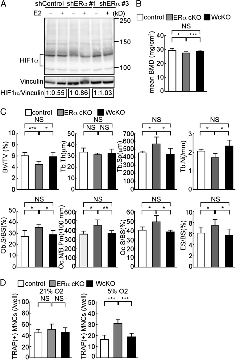
Fig. 3.
HIF1α deletion rescues bone phenotypes seen in ERα cKO mice in vivo. (A) Raw264.7 cells transfected with shRNA targeting ERα (shERα) or nontarget control shRNA (shControl) and treated with (+) or without (−) E2. HIF1α protein levels determined by immunoblot were quantified by densitometry and are shown as values relative to E2(−). (B) BMD of femurs divided equally longitudinally from control (CtskCre/+), ERα cKO (CtskCre/+; ERαf/f), and WcKO (CtskCre/+; Hif1αf/f; ERαf/f) mice. (C) Bone histomorphometrical analysis of femurs from control, ERα cKO, and WcKO female mice. (D) Osteoclast formation, as evidenced by TRAP positivity, of osteoclast progenitor cells from control, ERα cKO, and WcKO mouse bone marrow under normoxic (21%) or hypoxic (5%) conditions during the course of RANKL-stimulated differentiation. Data (B–D) represent the mean value of the indicated parameter ± SD (*P < 0.05; ***P < 0.005; NS, not significant; n = 5). White, gray, and black bars represent control, ERα cKO, and WcKO mice, respectively.

