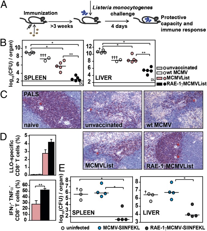
Fig. 2.
RAE-1γMCMV expressing antigenic peptides protects mice against L. monocytogenes. (A) Mice were immunized via f.p. with 105 pfu per mouse of WT MCMV or MCMV vector with or without RAE-1γ or were left nonimmunized. At 3 wk p.i. or later, mice were challenged with L. monocytogenes. On day 4 postchallenge, mice were sacrified and analyzed for an immune response and protective capacity to L. monocytogenes. (B) BALB/c mice were challenged 4 wk postimmunization with 2 × 10⁴ cfu per mouse of L. monocytogenes. Bacterial load was determined in spleen and liver. Individual animals (circles) and median values are shown. † indicates the death of a mouse. (C) Three weeks postimmunization, BALB/c mice were challenged with 104 cfu per mouse of L. monocytogenes. Paraffin-embedded spleen sections were stained for CD3ε expression. * indicates central artery. PALS, periarteriolar lymphoid sheath. (Magnification: 20×.) (D) Splenocytes were isolated and either stained with LLO-tetramers or stimulated with LLO-peptides and intracellularly stained for cytokine production (mean ± SEM, n = 4–5). (E) Three weeks postvaccination, C57BL/6 mice were challenged with 5 × 10⁴ cfu per mouse of OVA-Listeria. Individual animals and median values are shown.

