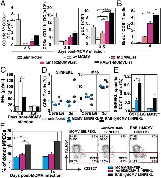
Fig. 6.
RAE-1γ expression promotes epitope-specific CD8 T cells priming. BALB/c mice were i.v. infected with 2 × 10⁵ pfu, and the following parameters were analyzed: (A) the frequency of DC subsets in spleen, (B) the frequency of proliferating CD8 T cells, and (C) IFN-α level in the sera. For A–C, mean ± SEM is shown; n = 3–5. (D) C57BL/6 and 3d mice were i.p. infected with 2 × 10⁵ pfu of indicated viruses and the specific CD8 T-cell response in spleen was determined on day 7 p.i. (E) C57BL/6 and Batf3−/− mice were infected i.v. with 2 × 10⁵ pfu of indicated viruses and SIINFEKL-specific CD8 T cells in spleen were determined on day 8 p.i. Mean ± SEM is shown. (F) Naive recipients (CD45.1) were transferred with 104 NKG2DΔ/ΔOT1 cells (CD45.2) and infected with 104 pfu per mouse of indicated viruses 24 h later. On days 7 and 14 p.i., donor CD8 T cells (CD45.1) were analyzed for the frequency of MPECs (KLRG1−CD127+ CD8 T cells) in blood (n = 3). Representative FACS plots of donor MPECs expansion are shown; n = 3–4.

