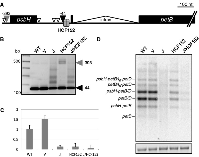
Figure 3.
Analysis of petB mRNA. (A) Schematic of the major bands detected by 5′ RACE. The filled gray and black arrowheads correspond to the similarly-labeled bands in (B). Open arrowheads represent estimated 5′ ends from products of intermediate sizes. HCF152, conserved minimal HCF152 binding site (15). (B) 5′ RACE products from RT priming within the petB exon. The filled black arrowhead marks the mature WT petB 5′ end (25) and the filled gray arrowhead marks products from a precursor transcript that terminates upstream of psbH, corresponding to the psbH-petB dicistron. Numbers at right are nucleotide positions relative to the petB translation initiation codon. (C) Relative amounts of petB-containing RNA fragments measured by qRT-PCR. (D) Strand-specific RNA gel blot for the petB exon. IB indicates unspliced petB species. An ethidium bromide-stained image of 28S rRNA is shown below to reflect loading.

