Figure 4.
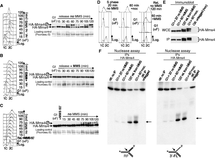
Mus81-Mms4 activation is not induced by DNA damage and occurs when cells finish S-phase. (A) HA-MMS4 cells (YMV33 strain) were synchronized in G1 and then released into fresh medium. Cell cycle progression was monitored by flow cytometry (left). Mms4 phosphorylation was analysed by immunoblot (right). (B) HA-MMS4 cells were blocked in G1 and then released into medium with MMS (0.02%). Left: Flow cytometry. Right: Immunoblot analysis of Mms4. (C) HA-MMS4 cells were synchronized in G1 and then released into S-phase in medium with MMS (0.02%) for 60 min. The MMS was then removed and the cells were allowed to progress through S-phase. Left: Flow cytometry. Right: Immunoblot analysis of Mms4. (D) Cell cycle experiments for the nuclease assays. HA-MMS4 cells were blocked in G1 and the culture was then divided in three. One culture was released in medium without MMS for 20 min; another one in medium without MMS plus nocodazole, for 60 min; the third one was released into S-phase for 60 min in medium with MMS (0.02%). The MMS was then removed and the cells were allowed to progress through the cell cycle. Cell cycle progression was followed by flow cytometry. (E) The extracts were prepared from cells taken at the indicated time points and HA-Mms4 was immunoaffinity purified. Mms4 was analysed by immunoblot in the whole cell extracts (WCE) and the yield of the IP was estimated. About 2% of the total amount of the IP protein used for the nuclease assays was loaded. (F) The nuclease activity was assayed by the resolution of a model RF (left panel) and a 3′-FL (right panel). An arrow indicates the labelled product resulting from the cleavage of each substrate. The controls were a reaction without extract and a nuclease assay using IP-extracts from untagged cells blocked in G2/M.

