Figure 1.
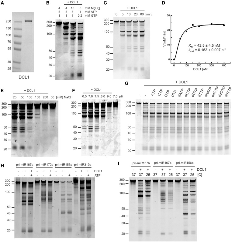
Characterization of pri-miRNA processing by DCL1. (A) Purified recombinant DCL1 protein. Coomassie blue-stained SDS–PAGE of purified DCL1 expressed in a baculovirus/insect cell expression system. (B) pri-miR167b processing by DCL1. The reaction was carried out under the same conditions as described previously (29) and at different concentrations of MgCl2, ATP and GTP. Pri-miR167b (150 nM) and DCL1 (50 nM) were incubated in a buffer [20 mM Tris–HCl (pH 7.0), 50 mM NaCl and indicated concentrations of MgCl2, ATP and GTP] at 37°C for 60 min. RNA was fractionated on 12% polyacrylamide/7.5 M urea denaturing gel and visualized by SYBR Gold staining. (C) Time course analysis of pri-miR167b processing. Pri-miR167b (150 nM) and DCL1 (50 nM) were incubated in a buffer [20 mM Tris–HCl (pH 7.0), 50 mM NaCl, 5 mM MgCl2 and 1 mM ATP] at 37°C for the indicated times. RNA was fractionated and detected as in (B). (D) pri-miR167b processing with a varying concentration of DCL1. The reaction was done at 37°C for 7 min with the same buffer components as in (C). The RNA was fractionated and detected as in (B). The amount of small RNA with 21mer and 23mer lengths was determined based on a series of known amounts of 20mer RNA fractionated alongside, and the velocity was plotted as a function of DCL1 concentration. (E) Effect of NaCl concentration. Pri-miR167b (150 nM) and DCL1 (50 nM) were incubated in a reaction buffer [20 mM Tris–HCl (pH 7.0), 5 mM MgCl2, 1 mM ATP and the indicated concentrations of NaCl] at 37°C for 20 min. The RNA was fractionated and detected as in (B). (F) Effect of pH. Pri-miR167b (150 nM) and DCL1 (50 nM) were incubated in a reaction buffer (50 mM NaCl, 5 mM MgCl2, 1 mM ATP and 20 mM Tris–HCl at the indicated pH) at 37°C for 20 min. The RNA was fractionated and detected as in (B). (G) Effect of nucleoside triphosphate. Pri-miR167b (150 nM) and DCL1 (50 nM) were incubated in a reaction buffer [20 mM Tris–HCl (pH 7.0), 50 mM NaCl, 5 mM MgCl2, and the indicated nucleoside triphosphates at 1 mM] at 37°C for 60 min. The RNA was fractionated and detected as in (B). (H) Effect of ATP on four pri-miRNA substrates. The reaction was done with the indicated pri-miRNA (150 nM) and DCL1 (50 nM) with or without 1 mM ATP as in (G). The RNA was fractionated and detected as in (B). (I) Effect of temperature. The reaction was done with the indicated pri-miRNA (150 nM) and DCL1 (50 nM) in a reaction buffer [20 mM Tris–HCl (pH 7.0), 5 mM MgCl2, 1 mM ATP and 50 mM NaCl] at 37°C or 25°C for 20 min. The RNA was fractionated and detected as in (B).

