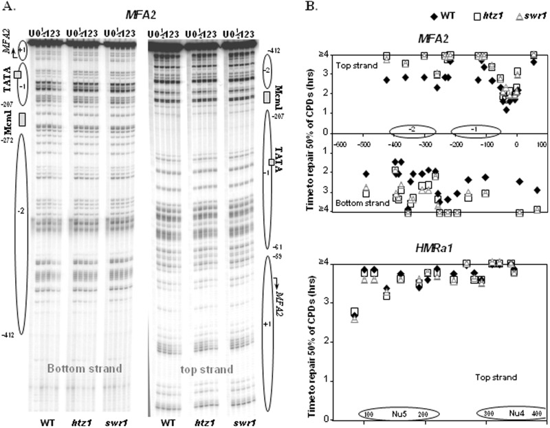
Figure 4.
Deletion of HTZ1 or SWR1 reduces the repair of CPDs in the MFA2 promoter. (A) Gels depicting CPDs in the top and bottom strands of a HaeIII restriction fragment (−516 to +83) in the MFA2 promoter after a UV dose of 100 J/m2. Lane U is DNA from mock-irradiated cells, while 0, 1/2, 1, 2 and 3 are DNA from irradiated cells after 0, 1/2, 1, 2 and 3 h repair, respectively. Alongside the gels are symbols representing nucleosome positions at MFA2. Nucleotide positions are allocated in relation to the MFA2 start codon. (B) Time to remove 50% of the initial CPDs (T50%) at given sites. T50% of a single CPD or a group of CPDs with a similar repair rate was calculated (T50% < 3 h) or extrapolated (T50% > 3 h). The T50% of slowly repaired CPDs (≥4 h) was shown at the same level (≥4 h) on the graph. Data are the average of three to five independent experiments.

