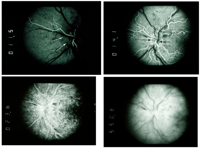
FIGURE 17.
Fluorescein angiography in diabetic papillopathy with prominent surface vascular dilation. Upper left, Fluorescein angiogram, early arteriovenous phase, showing absent optic disc filling (arrows) at 11.5 seconds. Upper right, Fluorescein angiogram, early arteriovenous phase, showing poor optic disc filling (arrows) and very early leakage from adjacent surface vessels at 14.1 seconds. Lower left, Fluorescein angiogram, mid phase, showing diffuse early leakage from dilated surface vessels at 23.6 seconds. Prelaminar layer of disc filling is obscured by dilated leaking surface vasculature. Lower right, Fluorescein angiogram, late phase, showing late leakage from optic disc at 550.8 seconds.

