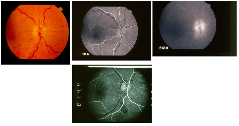
FIGURE 5.
Fluorescein angiography in nonarteritic anterior ischemic optic neuropathy diffuse. Upper left, Fundus photograph showing diffuse optic disc edema. Upper middle, Fluorescein angiogram, early arteriovenous phase, showing diffusely poor filling of optic disc (arrows) at 18.4 seconds. Upper right, Fluorescein angiogram, late phase, showing late leakage from optic disc at 613.8 seconds. Lower, Fluorescein angiography, early arteriovenous phase in normal eye, showing normal diffuse filling of the optic disc at 14.4 seconds

