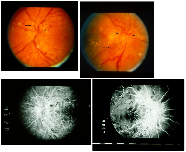
FIGURE 9.
Fluorescein angiography, diabetic papillopathy vs optic disc neovascularization. Upper left, Diabetic papillopathy, fundus photograph, optic disc edema with prominent surface vascular dilation. Vessels (arrows) predominantly are radial and extend minimally past disc margin. Upper right, Diabetic papillopathy and optic disc neovascularization, fundus photograph, optic disc edema with optic disc neovascularization. Vessels (arrows) are nonradial, irregular, and extend far past disc margin. Lower left, Diabetic papillopathy, fluorescein angiogram, mid phase, optic disc edema with prominent surface vascular dilation; leakage is primarily from radial disc surface vessels extending adjacent to disc. Lower right, Diabetic papillopathy and optic disc neovascularization, fluorescein angiogram, mid phase, optic disc edema with optic disc neovascularization, leakage from disc is relatively mild compared with extensive irregular vascular abnormality and leakage throughout posterior pole.

