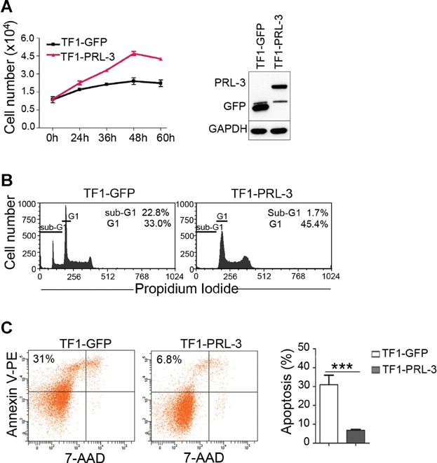
Figure 5. PRL-3 promotes growth and suppresses apoptosis of TF-1 leukaemia cells upon cytokine deprivation.

- Right panel: MTS assay results reflecting numbers of viable TF1-GFP and TF1-PRL-3 cells after culture in the absence of cytokines for various durations. Error bars represent the mean ± SD from three independent experiments. Left panel: Western blot analysis of TF1-GFP and TF1-PRL-3 cells. GAPDH, loading control.
- Flow cytometry analysis of propidium iodide-stained TF1-GFP and TF1-PRL-3 after 48 h culture in the absence of cytokines. Note the difference in sub-G1 peak/population, reflective of apoptotic cells. Representative data from three independent experiments are shown.
- Left panel: flow cytometry analysis of annexin-V- and 7-AAD-stained TF1-GFP and TF1-PRL-3 after 48 h culture in the absence of cytokines. The percentage in the upper left quadrant indicates the fraction of annexin-V-positive apoptotic cells in the entire cell population analysed. Right panel: quantitation of annexin-V-positive apoptotic population in three independent experiments. Statistical differences between two groups were determined using Student's t-test (mean ± SD, n = 3, ***p = 0.0012).
